|
Overview
 Fallen arches, or flatfoot, is a condition in which the arch on the inside of the foot is flat and the entire sole of the foot rests on the ground. It affects about 40% of the general population. Although flat feet in themselves are not usually problematic, they can create problems in the feet, hips, ankles and knees. Pain may be experienced in the lower back if there are alignment problems and if the individual is engaged in a lot of heavy, high impact activities that put stress on the bones and muscles in the lower legs. The arches of most individuals are fully developed by the age of 12 to 13. While some people are born with flat arches, for others the arches fall over time. The tibial tendon, which runs along the inside of the ankle from above the ankle to the arch, can weaken with age and with heavy activity. The posterior tendon, main support structure for the arch, can become inflamed (tendonitis) or even tear if overloaded. For women, wearing high heels can affect the Achilles tendon and alter the structure and function of the ankle. The posterior tibial tendon may compensate for this stress and break down, causing the arches to fall. Obesity is another contributing factor, as well as a serious injury to the ankle or foot, arthritis and bad circulation such as occurs with diabetes. Causes Family history, experts say fallen arches can run in families. Weak arch, the arch of the foot may be there when no weight is placed on it, for example, when the person is sitting. But as soon as they stand up the foot flattens (falls) onto the ground. Injury, arthritis, tibialis posterior (ruptured tendon), pregnancy, nervous system or muscle diseases, such as cerebral palsy, muscular dystrophy, or spina bifida. Tarsal Coalition, the bones of the foot fuse together in an unusual way, resulting in stiff and flat feet. Most commonly diagnosed during childhood. Diabetes. Age and wear and tear, years of using your feet to walk, run, and jump eventually may take its toll. One of the eventual consequences could be fallen arches. The posterior tibial tendon may become weakened after long-term wear a tear. The postario tibial tendon is the main support structure of the arch of our feet. The tendon can become inflamed (tendinitis) after overuse - sometimes it can even become torn. Once the tendon is damaged, the arch shape of the foot may flatten. Symptoms Knee/Hip/Back Pain - When the arch collapses in the foot, it triggers a series of compensations up the joint chain, leading to increased stress on the knee, pelvis and low back. Plantar fasciitis - This condition is characterized by heel pain, especially with the first few steps you take. The plantar fascia stretches as the arch falls, putting stress on the heel. Bunions - If you see a bony bump developing at the base of your big toe, you are likely developing a bunion. It may be swollen, red or painful when it rubs against your shoe. A flattened arch spreads the forefoot and causes the big toe to deviate toward the second toe. Shin splints - This term generally refers to pain anywhere along the shinbone. It is typically due to overuse and is aggravated after exercise and activity. Diagnosis Podiatrists are trained in expertly assessing flat feet and identifying different risk factors and the causes for it. Initial assessment will begin with a detailed history attempting to find out if any underlying illness has resulted in this. A detailed clinical examination normally follows. The patient may be asked to perform certain movements such as walking or standing on their toes to assess the function of the foot. Footwear will also be analysed to see if there has been excessive wear or if they are contributing to the pronation of the foot. To assess the structure of the foot further, the podiatrist may perform certain x-rays to get a detailed idea of the way the bones are arranged and how the muscle tissues may be affecting them. It also helps assess any potential birth defects in a bit more detail. Why do arches fall? Non Surgical Treatment If you have flat feet (pes planus) it does not mean you will have problems or pain. Flat feet are a common condition. Flat feet are defined as an abnormal lowering of the medial longitudinal arch. There is often a hereditary component to the development of flat feet. Flat feet are more common in certain ethnic groups. Flat feet usually occurs equally in both feet and become apparent at an early age. Flat feet can initially be asymptomatic, but can become progressively symptomatic with age. Some flat feet never become bothersome. The medial arch is unstable in flexible flatfoot. The foot can bend as the heel lifts of the ground. This creates stress on the surrounding soft-tissues and joints creating tired, sore arches. The instability of the foot can create abnormal stress that flows up the leg which results in pain in the legs, knees, hips and lower back. Patients with flat feet often present to the chiropodist/podiatrist with problems that are indirectly caused by their foot structure. Surgical Treatment  Fallen arches may occur with deformities of the foot bones. Tarsal coalition is a congenital condition in which the bones of the foot do not separate from one another during development in the womb. A child with tarsal coalition exhibits a rigid flat foot, which can be painful, notes the patient information website eOrthopod. Surgery may prove necessary to separate the bones. Other foot and ankle conditions that cause fallen arches may also require surgery if noninvasive treatments fail to alleviate pain and restore normal function. After Care Time off work depends on the type of work as well as the surgical procedures performed. . A patient will be required to be non-weight bearing in a cast or splint and use crutches for four to twelve weeks. Usually a patient can return to work in one to two weeks if they are able to work while seated. If a person's job requires standing and walking, return to work may take several weeks. Complete recovery may take six months to a full year. Complications can occur as with all surgeries, but are minimized by strictly following your surgeon's post-operative instructions. The main complications include infection, bone that is slow to heal or does not heal, progression or reoccurrence of deformity, a stiff foot, and the need for further surgery. Many of the above complications can be avoided by only putting weight on the operative foot when allowed by your surgeon. Overview
People with leg length discrepancy, when one leg is longer than the other, usually have a waddling-type gait where the hips seem to move up and down during walking as the body tries to compensate for the inequality. There are two types of leg length discrepancy. The first type of leg length discrepancy involves a structural defect, where one bone is longer or shorter than the corresponding bone of the other limb. This can occur within the femur (upper leg bone) or the tibia and fibular (lower leg bones). Functional leg length discrepancy results from altered mechanics due to a malalignment in the spine or lower extremity.  Causes A number of causes may lead to leg length discrepancy in children. Differences in leg length frequently follow fractures in the lower extremities in children due to over or under stimulation of the growth plates in the broken leg. Leg length discrepancy may also be caused by a congenital abnormality associated with a condition called hemihypertrophy. Or it may result from neuromuscular diseases such as polio and cerebral palsy. Many times, no cause can be identified. A small leg length discrepancy of a quarter of an inch or less is quite common in the general population and of no clinical significance. Larger leg length discrepancies become more significant. The long-term consequences of a short leg may include knee pain, back pain, and abnormal gait or limp. Symptoms The effects vary from patient to patient, depending on the cause of the discrepancy and the magnitude of the difference. Differences of 3 1/2 to 4 percent of the total length of the lower extremity (4 cm or 1 2/3 inches in an average adult), including the thigh, lower leg and foot, may cause noticeable abnormalities while walking and require more effort to walk. Differences between the lengths of the upper extremities cause few problems unless the difference is so great that it becomes difficult to hold objects or perform chores with both hands. You and your physician can decide what is right for you after discussing the causes, treatment options and risks and benefits of limb lengthening, including no treatment at all. Although an LLD may be detected on a screening examination for curvature of the spine (scoliosis), LLD does not cause scoliosis. There is controversy about the effect of LLD on the spine. Some studies indicate that people with an LLD have a greater incidence of low back pain and an increased susceptibility to injuries, but other studies refute this relationship. Diagnosis The doctor carefully examines the child. He or she checks to be sure the legs are actually different lengths. This is because problems with the hip (such as a loose joint) or back (scoliosis) can make the child appear to have one shorter leg, even though the legs are the same length. An X-ray of the child?s legs is taken. During the X-ray, a long ruler is put in the image so an accurate measurement of each leg bone can be taken. If an underlying cause of the discrepancy is suspected, tests are done to rule it out. Non Surgical Treatment Treatments for limb-length discrepancies and differences vary, depending on the cause and severity of the condition. At Gillette, our orthopedic surgeons are experts in typical and atypical growth and development. Our expertise lets us plan treatments that offer a lifetime of benefits. Treatments might include monitoring growth and development, providing noninvasive treatments or therapy, and providing a combination of orthopedic surgical procedures. To date, alternative treatments (such as chiropractic care or physical therapy) have not measurably altered the progression of or improved limb-length conditions. However, children often have physical or occupational therapy to address related conditions, such as muscle weakness or inflexibility, or to speed recovery following a surgical procedure. In cases where surgical treatment isn?t necessary, our orthopedists may monitor patients and plan noninvasive treatments, such as, occupational therapy, orthoses (braces) and shoe inserts, physical therapy, prostheses (artificial limbs).  shoe lifts for women's shoes Surgical Treatment Large leg length inequalities can be treated by staged lengthenings or by simultaneous ipsilateral femoral and tibial lengthenings. Additionally, lengthenings can be combined with appropriately timed epiphysiodesis in an effort to produce leg length equality. Staged lengthenings are often used for congenital deficiencies such as fibular hemimelia, in which 15 cm or more may be needed to produce leg length equality. We typically plan for the final lengthening to be completed by age 13 or 14 years, and allow at least 3 years between lengthenings. Lengthening of both the tibia and femur simultaneously requires aggressive therapy and treatment of soft tissue contractures. Curran et al[57] reported the need for surgical release of soft tissue contractures in 3 of 8 patients treated with simultaneous ipsilateral femoral and tibial lengthenings. Lengthening over an IM nail can be done in an effort to decrease the amount of time the fixator needs to be worn and to prevent angular malalignment. This technique requires that the patient be skeletally mature and it carries a higher risk of osteomyelitis (up to 15%). Additionally, if premature consolidation occurs, a repeat corticotomy is more difficult. Overview
 A Morton's neuroma is a benign (noncancerous) swelling along a nerve in the foot that carries sensations from the toes. The reason the nerve starts to swell is unknown. But once swelling begins, the nearby bones and ligaments put pressure on the nerve, causing more irritation and inflammation. This produces burning pain, numbness, tingling and other abnormal sensations in the toes. A Morton's neuroma also is called an interdigital neuroma, intermetatarsal neuroma or a forefoot neuroma. A Morton's neuroma is a benign (noncancerous) swelling along a nerve in the foot that carries sensations from the toes. The reason the nerve starts to swell is unknown. But once swelling begins, the nearby bones and ligaments put pressure on the nerve, causing more irritation and inflammation. This produces burning pain, numbness, tingling and other abnormal sensations in the toes. A Morton's neuroma also is called an interdigital neuroma, intermetatarsal neuroma or a forefoot neuroma.Causes Although the exact cause for this condition is unclear, a number of factors can contribute to the formation of a neuroma. Biomechanical deformities, such as a high-arched foot or a flat foot, can lead to the formation of a neuroma. These foot types bring on instability around the toe joints, leading to the development of the condition. Trauma can cause damage to the nerve, resulting in inflammation or swelling of the nerve. Improper footwear that causes the toes to be squeezed together is problematic. Avoid high-heeled shoes higher than two inches. Shoes at this height can increase pressure on the forefoot area. Repeated stress, common to many occupations, can create or aggravate a neuroma. Symptoms If you have a Morton's neuroma, you will probably have one or more of these symptoms. Tingling, burning, or numbness. A feeling that something is inside the ball of the foot, or your sock is bunched up. Pain that is relieved by removing your shoes. A Morton's Neuroma often develops gradually. At first the symptoms may occur only occasionally, when wearing narrower shoes or performing certain activities. The symptoms may go away temporarily by massaging the foot or by avoiding aggravating shoes or activities. Over time the symptoms progressively worsen and may persist for several days or weeks. The symptoms become more intense as the neuroma enlarges and the temporary changes in the nerve become permanent. Diagnosis Your health care provider can usually diagnose this problem by examining your foot. A foot x-ray may be done to rule out bone problems. MRI or high-resolution ultrasound can successfully diagnose Morton's neuroma. Nerve testing (electromyography) cannot diagnose Morton's neuroma, but may be used to rule out conditions that cause similar symptoms. Blood tests may be done to check for inflammation-related conditions, including certain forms of arthritis. Non Surgical Treatment You may need a metatarsal pad if wider shoes do not help relieve your Morton?s neuroma symptoms. A metatarsal pad will help spread your metatarsal bones and reduce pressure on your affected nerve as it travels under the ball of your foot. The placement of your metatarsal pad is important, and it is best placed by a foot care professional who has experience in the anatomy of the forefoot and Morton?s neuroma treatment.  Surgical Treatment The ultimate success of a Morton?s neuroma treated surgically can be variable. In cases where the underlying problem is only an irritated nerve (a true Morton?s neuroma), then surgery will probably be curative (although it may take a few months for the foot to fully heal). But in many cases, forefoot pain is more complex. There may be an irritated nerve or two causing pain, but the real problem is often excessive loading of the lesser metatarsals. The generic term for this condition is metatarsalgia. When considering surgery, identifying and addressing these problems may lead to a better end result. There are actually not one but two different kinds of leg length discrepancies, congenital and acquired. Congenital implies you are born with it. One leg is structurally shorter compared to the other. As a result of developmental periods of aging, the human brain senses the stride pattern and identifies some variance. The body usually adapts by dipping one shoulder over to the "short" side. A difference of under a quarter inch is not blatantly uncommon, require Shoe Lifts to compensate and generally does not have a profound effect over a lifetime.
 Leg length inequality goes typically undiagnosed on a daily basis, however this condition is easily solved, and can reduce a number of cases of back ache. Therapy for leg length inequality typically consists of Shoe Lifts . These are generally economical, often being under twenty dollars, in comparison to a custom orthotic of $200 or even more. Differences over a quarter inch can take their toll on the spine and should probably be compensated for with a heel lift. In some cases, the shortage can be so extreme that it requires a full lift to both the heel and sole of the shoe. Mid back pain is the most widespread condition impacting people today. Around 80 million men and women are affected by back pain at some stage in their life. It is a problem that costs businesses millions each year because of time lost and output. New and more effective treatment methods are constantly sought after in the hope of decreasing the economical influence this issue causes.  People from all corners of the earth experience foot ache due to leg length discrepancy. In a lot of these cases Shoe Lifts are usually of beneficial. The lifts are capable of relieving any pain in the feet. Shoe Lifts are recommended by numerous qualified orthopaedic practitioners". So that you can support the human body in a healthy and balanced manner, the feet have a significant part to play. Despite that, it is sometimes the most overlooked area of the human body. Some people have flat-feet meaning there may be unequal force placed on the feet. This causes other body parts such as knees, ankles and backs to be affected too. Shoe Lifts guarantee that suitable posture and balance are restored.  Overview Heel spurs are tiny protruding calcium deposits that can develop near the base of your heel bone. They can be caused by repetitive activities, such as dancing or running, or they can form in association with plantar fasciitis, which is an inflammation of the ligament (plantar fascia) on the bottom of your foot. When the plantar fascia is tight and pulls on your heel bone, the bone releases calcium to try to heal itself. The excess deposits of calcium can sometimes form heel spurs. Causes These bony protrusions are commonly found in association with a condition called Plantar Fasciitis. Plantar Fasciitis occurs when the plantar fascia ligament, which spans the arch of the foot, becomes stressed, torn and inflamed. Misalignment and excessive movement of the plantar fascia ligament is most typically the result of an abnormal walking gait. The ligament is designed to stretch with the bounce of each step you take, but if it stretches too much, the resulting small tears and inflammation can cause mild to extreme discomfort. At the same time, it is believed that heel spurs are formed as the body attempts to provide some additional support to the over-stretched ligament. In other words, calcium builds up as a prop to the plantar fascia. 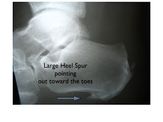 Symptoms With heel spurs, people often talk about a dull ache which is felt most of the time with episodes of a sharp pain in the center of the heel or on the inside margin of the heel. Often the pain is worse on first rising in the morning and after rest and is aggravated by prolonged weight bearing and thin-soled shoes. Diagnosis A Heel Spur diagnosis is made when an X-ray shows a hook of bone protruding from the bottom of the foot at the point where the plantar fascia is attached to the heel bone. The plantar fascia is the thick, connective tissue that runs from the calcaneus (heel bone) to the ball of the foot. This strong and tight tissue helps maintain the arch of the foot. It is also one of the major transmitters of weight across the foot as you walk or run. In other words, tremendous stress is placed on the plantar fascia. Non Surgical Treatment By reducing excessive motion and controlling and supporting the foot during physical activities an orthotic can help to limit how far the plantar fascia is pulled or torn away from the heel. A Heel Spur pad can be offered- which is a pad designed to take pressure off the spur. If the problem persists, consult your foot doctor. Surgical Treatment In a small number of cases (usually less than 5 percent), patients may not experience relief after trying the recommendations listed above. It is important that conservative treatments (such as those listed above) be performed for AT LEAST a year before considering surgery. Time is important in curing the pain from heel spurs, and insufficient treatment before surgery may subject you to potential complications from the procedure. If these treatments fail, your doctor may consider an operation to loosen the plantar fascia, called a plantar fascia release. Prevention To prevent this condition, wearing shoes with proper arches and support is very important. Proper stretching is always a necessity, especially when there is an increase in activities or a change in running technique. It is not recommended to attempt working through the pain, as this can change a mild case of heel spurs and plantar fascitis into a long lasting and painful episode of this condition.  Overview A bone spur (osteophyte) is a bony growth that forms along the edge of normal bone in response to wear and tear, most frequently in the joints. A heel spur is a bone spur of the heel bone, which causes heel pain by rubbing on the achilles tendon or other soft tissues. Causes The plantar fascia is a big strong ligament on the bottom of the foot, starting at the bottom of the heel bone and running into the ball of the foot. As the arch of the foot becomes weak, it sags slightly with each step and this causes the plantar fascia to tug and pull at the heel bone with each step. Over a period of time, a spur forms where this big strong ligament tugs and pulls at the heel bone. Soon, inflammation (swelling) starts around this spur and the pain becomes almost unbearable. (Sometimes heel spurs may be present without being painful if no inflammation is present).  Symptoms Some symptoms at the beginning of this condition include pain and swelling, and discomfort when pushing off with the toes during walking. This movement of the foot stretches the fascia that is already irritated and inflamed. If this condition is not treated, pain will be noticed in the heel when a heel spur develops in response to the stress. This is a common condition among athletes and others who run and jump a significant amount. Diagnosis Your doctor, when diagnosing and treating this condition will need an x-ray and sometimes a gait analysis to ascertain the exact cause of this condition. If you have pain in the bottom of your foot and you do not have diabetes or a vascular problem, some of the over-the-counter anti-inflammatory products such as Advil or Ibuprofin are helpful in eradicating the pain. Pain creams, such as Neuro-eze, BioFreeze & Boswella Cream can help to relieve pain and help increase circulation. Non Surgical Treatment Rest won?t help you in case of pain from the heel spur. When you get up after sleeping for some time, the pain may get worse. The pain worsens after a period of rest. You will feel pain because the plantar fascia elongates during working which stresses the heel. It is important to see a doctor if you are having consistent pain in you heel. The doctors may advise few or all of the conservative treatments, stretching exercises, shoe recommendations, shoe inserts or orthotic devices, physical therapy, taping or strapping to rest stressed muscles and tendons. There are some over-the-counter medicines available for treatment of heel pain. Acetaminophen (Tylenol), ibuprofen (Advil), or naproxen (Aleve) are some such medicines which can help you to get relief from the pain. In case of biomechanical imbalances causing the pain, a functional orthotic device can help you to get relief. Your doctor may also advise a corticosteroid injection for eliminating the inflammation. Surgical Treatment Sometimes bone spurs can be surgically removed or an operation to loosen the fascia, called a plantar fascia release can be performed. This surgery is about 80 percent effective in the small group of individuals who do not have relief with conservative treatment, but symptoms may return if preventative measures (wearing proper footwear, shoe inserts, stretching, etc) are not maintained. Prevention The best way to prevent heel spurs is by wearing properly fitted footwear. Shoes should have a shock absorbing tread and soles and should be effective in supporting the heel and arch. Proper warm up and stretching before embarking on any physical activity that will put pressure or impact on the area is highly recommended. Also, just as it?s important for your general health, if you can lose some extra pounds, you will be more likely to avoid heel spurs. If you are starting to feel the onset of pain, it may not be heel spurs, but could be a tendonitis condition that could lead to heel spurs. Overview
Calcaneal bursitis is seen somewhat frequently, particularly if the clinician has a predominantly musculoskeletal practice. No mortality is associated with calcaneal bursitis. Morbidity is associated with progressive pain and limping (antalgic gait) in patients who have not received adequate treatment. If chronic inflammation also affects the distal Achilles tendon, rupture of the tendon may occur. No race predilection has been documented. Calcaneal bursitis is observed in men and women. However, some increased risk may be incurred by women who wear high-heeled shoes. Calcaneal bursitis is commonly observed in middle-aged and elderly persons, the condition is also seen in athletes of all ages.Bursitis is a painful swelling of a small sac of fluid called a bursa. Bursae (plural of bursa) cushion and lubricate areas where tendons, ligaments, skin, muscles, or bones rub against each other. People who repeat the same movement over and over or who put continued pressure on a joint in their jobs, sports, or daily activities have a greater chance of getting bursitis. Causes Feet are extremely resilient and are designed to stand up to the pressures of day-to-day living. In some cases, though, foot structures may break down when subjected to chronic stress associated with long periods of weight-bearing activity on concrete, asphalt, or other hard surfaces (especially when footwear does not allow for proper weight distribution). Foot problems, including infracalcaneal bursitis, are often made worse by poorly designed footwear, and pressure, impact, and shear forces can damage the feet over time. Bursal sacs are intended to minimize this damage, but sometimes the bursa itself becomes inflamed. A rapid increase in physical activity levels or thinning of the heel?s protective fat pad are factors that may contribute to infracalcaneal bursitis. Other possible causes of infracalcaneal bursitis include blunt force trauma. Arthritic conditions. Acute or chronic infection. The following factors may increase a person?s risk of bursitis, including infracalcaneal bursitis. Poor conditioning. Exposure to cold weather. Participating in contact sports. Having a previous history of bursitis in any joint. Heel striking when running, especially in conventional running shoes with heel elevation. Symptoms The main symptom of heel bursitis is pain. You may experience pain in your heel when you walk or run. There may also be pain if the area is touched or if you stand on your tiptoes. In addition to pain, the area may appear red and warm, which are both signs of inflammation. Even if you have these symptoms, only a doctor can determine if you have bursitis of the heel. Your doctor will use these symptoms along with a general exam to determine if you are suffering from bursitis of the heel. Diagnosis Your doctor will examine you, including an evaluation of your gait, while you are barefoot, your doctor will ask you to stand still and to walk in order to evaluate how your foot moves as you walk. An examination of your feet. Your doctor may compare your feet for any differences between them. Then your doctor may examine your painful foot for signs of tenderness, swelling, discoloration, muscle weakness and decreased range of motion. A neurological examination. The nerves and muscles may be evaluated by checking strength, sensation and reflexes. In addition to examining you, your health care professional may want to examine your shoes. Signs of excessive wear in certain parts of a shoe can provide valuable clues to problems in the way you walk and poor bone alignment. Depending on the results of your physical examination, you may need foot X-rays or other diagnostic tests. Non Surgical Treatment All types of bursitis often can be successfully managed non-surgically, and possible treatments include use of ice packs or compressive dressings, activity modification that may reduce stress or irritation, administration of nonsteroidal anti-inflammatory drugs (NSAIDs) or antibiotics, corticosteroid injections (knee and elbow), stretching exercises, and/or change of footwear (heel). Surgery may be required in patients whose symptoms remain following these treatments and in certain situations when infection is involved. Surgical Treatment Bursectomy is a surgical procedure used to remove an inflamed or infected bursa, which is a fluid-filled sac that reduces friction between tissues of the body. Because retrocalcaneal bursitis can cause chronic inflammation, pain and discomfort, bursectomy may be used as a treatment for the condition when it is persistent and cannot be relived with other treatments. During this procedure, a surgeon makes small incisions so that a camera may be inserted into the joint. This camera is called an arthroscope. Another small incision is made so that surgical instruments can be inserted to remove the inflamed bursa.  Overview OverviewHammer toes most commonly affects the second toe on the foot. It causes the middle joint to bend. Hammertoe is most frequently caused by structural problems in the toe or from wearing poor fitting shoes. It is important to diagnose and treat hammertoe early because the condition tends to become worse over time. If left untreated, hammertoe can require surgery. Causes Hammer toe may also be caused by other medical conditions such as rheumatoid arthritis, osteoarthritis, or stroke because these forms of illnesses involve affectation of the person's muscles and nerves. Diabetes is also a causative factor for hammer toes due to diabetic neuropathy, which often times accompanies advanced instances of diabetes. Injury to a person's toes may also cause hammer toes, particularly if the injury involves breaking of the toes. In some instances, hammer toes may be hereditary. Some people may be genetically predisposed to develop the condition because of the natural structure of their bodies.  Symptoms SymptomsSymptoms of a hammertoe are usually first noticed as a corn on the top of the toe or at the tip which produces pain with walking or wearing tight shoes. Most people feel a corn is due to a skin problem on their toes, which in fact, it is protecting the underlying bone deformity. A corn on the toe is sometimes referred to as a heloma dura or heloma durum, meaning hard corn. This is most common Hammer toe at the level of the affected joint due to continuous friction of the deformity against your shoes. Diagnosis Hammertoes are progressive, they don?t go away by themselves and usually they will get worse over time. However, not all cases are alike, some hammertoes progress more rapidly than others. Once your foot and ankle surgeon has evaluated your hammertoes, a treatment plan can be developed that is suited to your needs. Non Surgical Treatment Podiatric Care may include using anti-inflammatory oral medications or an injection of medication and local anesthetic to reduce this swelling. When you go to your doctor, x-rays are usually required to evaluate the structure of your foot, check for fractures and determine the cause. The podiatrist may see you to take care of any corns that develop due to the bone deformities. They may advise you on different shoewear or prescribe a custom made orthotic to try and control the foot structure. Padding techniques may be used to straighten the toe if the deformity is flexible, or pads may be used to lessen the pressure on the area of the corn or ulcer. Your podiatric physician may also recommend a surgical procedure to actually fix the structural problem of your foot. Surgical Treatment Toes can be surgically realigned and made straight again. They can even be made shorter. The good news is that toes can be corrected. Hammer toe surgery is often synonymous with ?toe shortening?, ?toe job? and/or ?toe augmentation?. Depending on the severity and length of the toe, there are several methods to surgically correct a hammer toe. In general, the surgery involves removing a portion of the bone at the contracted joint, to realign the toe.  Overview OverviewA hammertoe is a deformity that causes your toe to bend or curl downward instead of pointing forward. This deformity can affect any toe on your foot; however, it most often affects the second toe or third toe. Although a hammertoe may be present at birth, it usually develops over time due to wearing ill-fitting shoes or arthritis. In most cases, a hammertoe is treatable. Causes Hammer toe most frequently results from wearing poorly fitting shoes that can force the toe into a bent position, such as excessively high heels or shoes that are too short or narrow for the foot. Having the toes bent for long periods of time can cause the muscles in them to shorten, resulting in the hammer toe deformity. This is often found in conjunction with bunions or other foot problem (e.g., a bunion can force the big toe to turn inward and push the other toes). It can also be caused by muscle, nerve, or joint damage resulting from conditions such as osteoarthritis, rheumatoid arthritis, stroke, Charcot-Marie-Tooth disease, complex regional pain syndrome or diabetes. Hammer toe can also be found in Friedreich's ataxia.  Symptoms SymptomsPain upon pressure at the top of the bent toe from footwear. The formation of corns on the top of the joint. Redness and swelling at the joint contracture. Restricted or painful motion of the toe joint. Pain in the ball of the foot at the base of the affected toe. Diagnosis Most health care professionals can diagnose hammertoe simply by examining your toes and feet. X-rays of the feet are not needed to diagnose hammertoe, but they may be useful to look for signs of some types of arthritis (such as rheumatoid arthritis) or other disorders that can cause hammertoe. If the deformed toe is very painful, your doctor may recommend that you have a fluid sample withdrawn from the joint with a needle so the fluid can be checked for signs of infection or gout (arthritis from crystal deposits). Non Surgical Treatment Conservative treatment starts with new shoes that have soft, roomy toe boxes. Shoes should be one-half inch longer than your longest toe. For many people, the second toe is longer than the big toe.) Avoid wearing tight, narrow, high-heeled shoes. You may also be able to find a shoe with a deep toe box that accommodates the hammer toe. Or, a shoe repair shop may be able to stretch the toe box so that it bulges out around the toe. Sandals may help, as long as they do not pinch or rub other areas of the foot. Surgical Treatment Hammer toe can be corrected by surgery if conservative measures fail. Usually, surgery is done on an outpatient basis with a local anesthetic. The actual procedure will depend on the type and extent of the deformity. After the surgery, there may be some stiffness, swelling and redness and the toe may be slightly longer or shorter than Hammer toes before. You will be able to walk, but should not plan any long hikes while the toe heals, and should keep your foot elevated as much as possible.  Prevention PreventionThe key to prevention is to wear shoes that fit you properly and provide plenty of room for your toes. Here?s how to get the right fit. Have your feet properly measured. The best way to do this is to get someone to draw the outline of your foot while you stand barefoot with your full weight on it, then measure the outline at the widest point. Measure the soles of your shoes. Ideally, they should be as wide as your feet, but certainly no more than half an inch narrower. Length matters, too, of course: your shoes should be half an inch longer than your longest toe. Overview
One of the main postural deviations that cause pain and injury in the foot and ankle area (and resultant compensations in the rest of the body) is overpronation. Pronation is a normal function that occurs when the foot rolls inward toward the midline of the body. This movement causes the heel to collapse inward and the medial arch of the foot to elongate and flatten. Overpronation, however, is when the foot collapses too far inward for normal function. Consequently, this directly affects the ability of the foot to perform and can disrupt proper functioning through the entire body.  Causes Pronation can occur as an overuse syndrome in active runners, where a great deal of stress is placed on ligaments and tendons that support the medial column. Obesity is another predictor for pronation and deterioration of the medial ligaments and posterior tibial tendon due to excessive stress on these tissues. Acute Trauma can also lead to over-pronation when ligaments are torn or tendon is ruptured. Once again this can lead to a collapse of the medial column. Arthritic conditions involving the knee joint when the joint is in varus (inner collapse) posture, this places the center of gravity over the ankle joint rather than the foot causing undue pressure on the inner ankle. Symptoms Overpronation can negatively affect overall body alignment. The lowering of the longitudinal arch pulls the heel bone in, causing the leg, thigh bone and hip to rotate inwards, and an anterior tilt of the pelvis. Unnecessary strain to the ankles, knees, hips and back can result. Plantar fasciitis and inflammation, metatarsal pain, problems with the Achilles tendon, pain on the inside of the knee, and bursitis in the hip are just some of the conditions commonly associated with pronation. Diagnosis Do the wet foot test. Get your feet wet and walk along a paved surface or sand and look at the footprints you leave. If you have neutral feet you will see a print of the heel with a thin strip connecting to your forefoot, but if you're overpronating your foot print will look a bit like a giant blob with toes.  Non Surgical Treatment If you overpronate, you should talk with a foot and ankle specialist, especially if symptoms have not developed yet. Questions you may want to ask your doctor include what are the best running shoes on the market? Where can I find those shoes? If over-the-counter orthotics don?t work, how long should I wait before contacting you for custom-made orthotics? On my next visit, what type of diagnostic testing should I expect? If I limit the amount of time I spend running, will my overpronation symptoms disappear? What additional treatment options can we try? Surgical Treatment Subtalar Arthroereisis. The ankle and hindfoot bones/midfoot bones around the joint are fused, locking the bones in place and preventing all joint motion. This may also be done in combination with fusion at other joints. This is a very aggressive option usually reserved for extreme cases where no joint flexibility is present and/or the patient has severe arthritic changes in the joint. |
