|
Overview
Sever's disease involves a lesion to the heel that appears mostly as a result of excessively twisting the ankle. The injury is particularly prevalent in children over 10 and other younger athletes because the heel bone, muscles and tendons are still developing - the bone faster than the muscles - and the condition strikes a growing part of the bone called the calcaneal apophysis. In older people an injury of this sort generally results in a stress fracture. Causes Sever?s disease is directly related to overuse of the bone and tendons in the heel. This can come from playing sports or anything that involves a lot of heel movement. It can be associated with starting a new sport, or the start of a new season. Children who are going through adolescence are also at risk of getting it because the heel bone grows quicker than the leg. Too much weight bearing on the heel can also cause it, as can excessive traction since the bones and tendons are still developing. It occurs more commonly in children who over-pronate, and involves both heels in more than half of patients. Symptoms This is a condition that affects the cartilage growth plate and the separate island of growing bone on the back of the heel bone. This growth plate is called the physeal plate. The island of growing bone is called the apophysis. It has the insertion attachment of the Achilles tendon, and the attachment of the plantar fascia. This island of bone is under traction from both of these soft tissue tendon and tendon-like attachments. Diagnosis Sever condition is diagnosed by detecting the characteristic symptoms and signs above in the older children, particularly boys between 8 and 15 years of age. Sometimes X-ray testing can be helpful as it can occasionally demonstrate irregularity of the calcaneus bone at the point where the Achilles tendon attaches. Non Surgical Treatment If your child have Sever's disease, the following is suggested, cut back on sporting activities, don't stop, just reduce the amount until symptoms improve (if the condition has been present for a while, a total break from sport may be needed later) avoid going barefoot, a soft cushioning heel raise is really important (this reduces the pull from the calf muscles on the growth plate and increases the shock absorption, so the growth plate is not knocked around as much). Stretch the calf muscles, provided the stretch does not cause pain in the area of the growth plate). The use of an ice pack after activity for 20mins is often useful for calcaneal apophysitis - this should be repeated 2 to 3 times a day. Recovery In some cases, children will simply outgrow Sever's Disease when they reach a certain age, but this does not mean that symptoms should be ignored. If children express that they are in pain, this should always be taken seriously by their parents or guardians. Heel pain may be a sign of Sever's Disease and this condition should not be left untreated, due to the damage it can cause to the growing heel bones. Scheduling a doctor's appointment is always the first step to take in gaining a diagnosis of symptoms and speedy help for the child. Overview
In growing children, legs can be made equal or nearly equal in length with a relatively simple surgical procedure. This procedure slows down the growth of the longer leg at one or two growth sites. Your physician can tell you how much equalization can be gained by this procedure. The procedure is performed under X-ray control through very small incisions in the knee area. This procedure will not cause an immediate correction in length. Instead, the limb length discrepancy will gradually decrease as the opposite extremity continues to grow and "catch up." Timing of the procedure is critical. The goal is to reach equal leg length by the time growth normally ends. This is usually in the mid-to-late teenage years. Disadvantages of this option include the possibility of slight over-correction or under-correction of the limb length discrepancy. In addition, the patient's adult height will be less than if the shorter leg had been lengthened. Correction of significant limb length discrepancy by this method may make a patient's body look slightly disproportionate because of the shorter leg. In some cases the longer leg can be shortened, but a major shortening may weaken the muscles of the leg. In the thighbone (femur), a maximum of 3 inches can be shortened. In the shinbone, a maximum of 2 inches can be shortened.  Causes Leg length discrepancies can be caused by: hip and knee replacements, lower limb injuries, bone diseases, neuromuscular issues and congenital problems. Although discrepancies of 2 cm or less are most common, discrepancies can be greater than 6 cm. People who have LLD tend to make up for the difference by over bending their longer leg or standing on the toes of their shorter leg. This compensation leads to an inefficient, up and down gait, which is quite tiring and over time can result in posture problems as well as pain in the back, hips, knees and ankles. Symptoms Patients with significant lower limb length discrepancies may walk with a limp, have the appearance of a curved spine (non-structural scoliosis), and experience back pain or fatigue. In addition, clothes may not fit right. Diagnosis A qualified musculoskeletal expert will first take a medical history and conduct a physical exam. Other tests may include X-rays, MRI, or CT scan to diagnose the root cause. Non Surgical Treatment The way in which we would treat a LLD would depend on whether we have an anatomical or functional difference. To determine which one is causing the LLD you will need to get your legs measured. This is the easiest way to determine if it is anatomical or functional. With a functional LLD we must first determine the cause and treat the cause. Should the cause be one that is not correctable then we may need to treat the LLD as if it were an anatomical or may have to treat the opposite leg to improve one's gait. As for the anatomical LLD, we may start off with a heel lift only in the shoe and follow up to see if we will need to put the lift full sole on the bottom of the shoe. This is determined by the affects that a heel lift in one shoe may have on that knee. Should the LLD be more than 1/4 inch we usually recommend starting between 1/8 inch to 1/4 inch less than the actual amount and let the body adjust to the change and then raise up to the measured amount later.  Surgical Treatment Differences of an inch-and-a-half to two inches may require epiphysiodesis (adjusting the growth of the longer side) or acute shortening of the other side. Differences greater than 2.5 inches usually require a lengthening procedure. The short bone is cut and an external device is applied. Gradual lengthening is done over months to allow the muscles and nerves accommodate the new length.
Overview
Adult flatfoot (adult acquired flatfoot) or posterior tibial tendon dysfunction (PTTD) is a common pathology presented to foot and ankle specialists. PTTD is characterized by a valgus (everted) hindfoot, flattening of the longitudinal arch of the foot (collapse) and abduction of the forefoot. This is a progressive deformity that begins flexible and can become rigid over time. The posterior tibial tendon (PT) is one of the main supporting structures of the foot arch. Changes within this tendon cause flattening of the foot. There are four stages of this deformity that begins flexible and progressives, with no treatment, to a rigid deformity and with time may involve the ankle joint. Patients usually present with pain in the foot or ankle stating the ?ankle? is rolling. It?s also common for patients to state they have difficulty walking barefoot. Pain is exacerbated after physical activities. Pain is usually isolated to the inside of the foot along the course of the PT tendon. 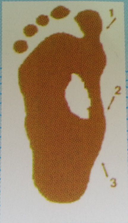 Causes Damage to the posterior tendon from overuse is the most common cause for adult acquired flatfoot. Running, walking, hiking, and climbing stairs are activities that add stress to this tendon, and this overuse can lead to damage. Obesity, previous ankle surgery or trauma, diabetes (Charcot foot), and rheumatoid arthritis are other common risk factors. Symptoms Not everyone with adult flatfoot has problems with pain. Those who do usually experience it around the ankle or in the heel. The pain is usually worse with activity, like walking or standing for extended periods. Sometimes, if the condition develops from arthritis in the foot, bony spurs along the top and side of the foot develop and make wearing shoes more painful. Diabetic patients need to watch for swelling or large lumps in the feet, as they may not notice any pain. They are also at higher risk for developing significant deformities from their flatfoot. Diagnosis Observe forefoot to hindfoot alignment. Do this with the patient sitting and the heel in neutral, and also with the patient standing. I like to put blocks under the forefoot with the heel in neutral to see how much forefoot correction is necessary to help hold the hindfoot position. One last note is to check all joints for stiffness. In cases of prolonged PTTD or coalition, rigid deformity is present and one must carefully check the joints of the midfoot and hindfoot for stiffness and arthritis in the surgical pre-planning. Non surgical Treatment Footwear has an important role, and patients should be encouraged to wear flat lace-up shoes, or even lace-up boots, which accommodate orthoses. Stage I patients may be able to manage with an off the shelf orthosis (such as an Orthaheel or Formthotics). They can try a laced canvas ankle brace before moving to a casted orthosis. The various casted, semirigid orthoses support the medial longitudinal arch of the foot and either hold the heel in a neutral alignment (stage I) or correct the outward bent heel to a neutral alignment (stage II). This approach is meant to serve several functions: to alleviate stress on the tibialis posterior; to make gait more efficient by holding the hindfoot fixed; and thirdly, to prevent progression of deformity. Devices available to do this are the orthosis of the University of California Biomechanics Laboratory, an ankle foot orthosis, or a removable boot. When this approach has been used, two thirds of patients have good to excellent results.  Surgical Treatment Stage two deformities are less responsive to conservative therapies that can be effective in mild deformities. Bone procedures are necessary at this stage in order to recreate the arch and stabilize the foot. These procedures include isolated fusion procedures, bone grafts, and/or the repositioning of bones through cuts called osteotomies. The realigned bones are generally held in place with screws, pins, plates, or staples while the bone heals. A tendon transfer may or may not be utilized depending on the condition of the posterior tibial tendon. Stage three deformities are better treated with surgical correction, in healthy patients. Patients that are unable to tolerate surgery or the prolonged healing period are better served with either arch supports known as orthotics or bracing such as the Richie Brace. Surgical correction at this stage usually requires fusion procedures such as a triple or double arthrodesis. This involves fusing the two or three major bones in the back of the foot together with screws or pins. The most common joints fused together are the subtalar joint, talonavicular joint, and the calcaneocuboid joint. By fusing the bones together the surgeon is able to correct structural deformity and alleviate arthritic pain. Tendon transfer procedures are usually not beneficial at this stage. Stage four deformities are treated similarly but with the addition of fusing the ankle joint. |
